Gastroenterological Procedures

Diagnostic Procedures

Oesophago-Gastro-Duodenoscopy
Otherwise known as Gastroscopy, this is an examination of the inside of the upper digestive system. It looks at the oesophagus, stomach and the first part of the small intestine called the duodenum. It is usually used to investigate symptoms such as difficulty in swallowing, heartburn, abdominal, nausea and vomiting. The procedure usually takes no longer than 15 minutes.
Colonoscopy
This procedure examines the large intestine or colon and can also examine the last part of the small intestine, called the terminal ileum, that joins the large intestine. The colonoscope is passed into your large intestine through your back passage. The examination usually takes 15 to 30 minutes, depending on how long, looped or kinked your large intestine is. This test is done to investigate bleeding from the back passage, changes in bowel habits, abdominal pain and to screen for polyps and colon cancer.
Enteroscopy of the small bowel
The more common examinations by Gastroscopy and Colonoscopy would be able to diagnose the majority of the problems of the digestive system. The small intestine is 6 m long. The first and second parts of the duodenum in the upper digestive tract as well as the terminal ileum that joins the colon are easily accessed. However the middle part of the small intestine is more difficult to examine. Balloon Enteroscopy is performed by a special endoscope which is fitted with a sheath which has an inflatable balloon at one end. By a process of inflating and deflating the balloon while moving the endoscope, the endoscope can reach from one end of the small intestine to the other end. The endoscope can be introduced either through the mouth or through the back passage, depending on where the problem is suspected to be. This method is used to investigate anaemia or bleeding, suspected tumours in the small intestine or inflammatory bowel disease, typically Crohn’s Disease.
Capsule Endoscopy of the small bowel
This endoscopic examination method is used to examine the bile ducts, which connect the liver to the small intestine and the pancreas. The endoscope is a side viewer called a Duodenoscope and is passed through the mouth, past the stomach and into the second part of the upper small intestine or duodenum. The Ampulla of Vater, which is the entrance to the bile and pancreatic ducts, is located and a catheter is used to enter the common bile duct or pancreatic duct. Contrast (a dye that is visible on x-ray) is injected and by Fluoroscopy (live, real-time x-ray scanning) these ducts can be examined. This examination is done if the bile ducts are thought to be blocked by gallstones or a tumour.
Endoscopic Retrograde Cholangio-Pancreatography (ERCP)

This endoscopic examination method is used to examine the bile ducts, which connect the liver to the small intestine and the pancreas. The endoscope is a side viewer called a Duodenoscope and is passed through the mouth, past the stomach and into the second part of the upper small intestine or duodenum. The Ampulla of Vater, which is the entrance to the bile and pancreatic ducts, is located and a catheter is used to enter the common bile duct or pancreatic duct. Contrast (a dye that is visible on x-ray) is injected and by Fluoroscopy (live, real-time x-ray scanning) these ducts can be examined. This examination is done if the bile ducts are thought to be blocked by gallstones or a tumour.

Liver Biopsy

Liver biopsy may be performed to diagnose inflammation, liver scarring (cirrhosis), cancerous or non-cancerous growths or some infections in the liver. Liver biopsy involves removing a tiny piece of liver tissue under a local anaesthetic, using a special liver biopsy needle. The specimen is then sent for pathological examination. Ultrasound scanning is commonly used to locate the best and safest place to insert the biopsy needle.

Therapeutic Procedures

By gastroscopy
Dilatation of oesophageal strictures
Narrowing (stricture) of the oesophagus may occur for a variety of reasons and causes difficulty in swallowing, or the sensation that food is “stuck” in the throat. These symptoms can be helped by a procedure in which the narrowed area of the oesophagus is “stretched” to widen it. A Gastroscope is first used to examine the narrowed segment of the oesophagus. Depending on the situation, a dilator may be in the form of a cylindrical balloon tightly wrapped onto a catheter which can be passed down the Gastroscope, across the narrowing, and the balloon inflated with water to stretch the narrowing. Some dilators require a guide wire to be put across the narrowing, the Gastroscope to be removed and a solid dilating device passed along the guide wire to dilate the oesophagus. It is sometimes necessary to use x-rays to guide this procedure.
Insertion of oesophageal stents
A narrowing of the oesophagus may be treated by inserting a tube or “stent”, made of plastic or metal, across the narrowed segment. Most modern stents are made of a mesh or lightweight “memory metal” that holds its final shape permanently. The stent holds the oesophagus open and improves swallowing. A stent is inserted using a Gastroscope. Sometimes the narrowed segment is first stretched or dilated and then the stent is placed into the oesophagus. There is no sensation of the stent being present and swallowing is often improved at once.
Injection sclerotherapy and band-ligation of oesophageal varices
Oesophageal varices are veins under the surface of the oesophagus that have become swollen due to liver scarring and increased pressure of blood flowing through the liver. As they increase in size there is an increased risk that they will bleed. To prevent major blood loss, the varices can be treated. Treatment is done through a Gastroscope and involves “shrinking” the varices. Injection sclerotherapy and variceal band-ligation are techniques use to shrink the oesophageal varices. In recent times, band-ligation has emerged as the favoured method with better efficacy and greater safety. Injection sclerotherapy involves injecting special medicines into the varices to cause them to inflame and shrink. Band-ligation of varices involves placing a small rubber band around the base of each swollen vein causing it to die and shrink. The band drops off and passes naturally through the digestive tract.
Insertion of percutaneous endoscopic gastrostomy (PEG) feeding tube
Some patients, due to illness such as stroke or physical obstruction such as cancer, are unable to take any food or nutrition by mouth. An alternate route of feeding such patients must be found. A PEG feeding tube is one such method, in which a feeding tube is inserted through the abdominal wall into the stomach and secured by retainers. Liquid food can be passed through the PEG tube directly into the stomach. No major surgery is required and insertion of a PEG tube is an endoscopic procedure. It is usually performed by two operators, one using an endoscope to examine the stomach and determine the best point of penetration and the other making a small incision in the skin of the abdominal wall, introducing a guide wire which is retrieved via the Gastroscope. The PEG tube is passed along the guide wire through the mouth and through the stomach wall to the outside where it is then secured.
Haemostasis (excluding oesophageal varices which is discussed elsewhere)
Bleeding can occur from a variety of lesions in the upper digestive tract. Typically it is due to gastric or duodenal ulcers. Other causes are abnormal blood vessels or tumours. Haemostasis means “stopping the bleeding”. If bleeding is brisk or there is evidence of recent significant bleeding from lesions, various means can be employed to stop the bleed. Injection needles can be passed down the Gastroscope and a diluted solution of Adrenaline can be injected around the bleeding point. This not only constricts blood vessels, but the effect of the liquid injected can compress the bleeding vessel (“tamponade”). In addition to this injection, a second method is often used and this can be a heater probe, Argon-plasma beam or electrical diathermy to coagulate the bleeding vessel or the use of special clips which can be deployed through the scope. In very profuse bleeding where the bleeding site might not even be easily seen, a powder called Haemospray can be sprayed onto the bleeding area. In contact with blood the powder coagulates and forms a shield that stops the bleeding and when the blood has cleared, a more precise method can be used later.
Laser or Argon-Plasma Beam re-canalisation
If inoperable cancers block the oesophagus, stomach or upper small intestine (duodenum), it is possible to cut the cancer back to enlarge the passageway. This can be done by either a Neodymium-Yttrium Aluminium Garnet (Nd-YAG) laser beam or an Argon-Plasma beam. The beams are delivered through fibres inserted through a Gastroscope and the tumour is incinerated piecemeal under direct vision.
By Colonoscopy
Removal of polyps
Colon polyps are often the precursors of colon cancers. When found they must be removed for examination to determine whether the cell type is potentially dangerous or whether there is already a focus of cancer inside the polyp. Polyps are of two broad kinds, pedunculated (with a neck) and sessile (no neck, sitting on the lining of the colon). Polyps can be removed by snare diathermy in which a catheter containing an extensible flexible metal loop is introduced into the colon via a Colonoscope and the polyp is “lassoed”. An electrical current is passed as the loop is closed, thus cutting the polyp off the lining of the colon. If it is a pedunculated polyp, cutting the polyp off at the neck is relatively simple. If the polyp has no neck, sometimes it is necessary to inject saline solution into the lining to raise a blister with the polyp on top, so that when snared off, there is a much reduced risk of perforating the colon.
Haemostasis (stopping bleeding) of bleeding lesions
Bleeding in the colon can come from Diverticulitis (inflamed out-pouches of the colonic wall), ulceration from infections or inflammatory bowel disease, from polyps, tumours and cancers or from haemorrhoids (piles – discussed elsewhere). Various means can be employed to stop the bleed. Injection needles can be passed down the Colonoscope and a diluted solution of Adrenaline can be injected around the bleeding point. This not only constricts blood vessels, but the effect of the liquid injected can compress the bleeding vessel (“tamponade”). In addition to this injection, a second method is often used and this can be a heater probe, Argon-plasma beam or electrical diathermy to coagulate the bleeding vessel or the use of special clips which can be deployed through the scope. In very profuse bleeding where the bleeding site might not even be easily seen, a powder called Haemospray can be sprayed onto the bleeding area. In contact with blood the powder coagulates and forms a shield that stops the bleeding and when the blood has cleared, a more precise method can be used later.
Insertion of stents
The colon can be blocked by either prior inflammation (such as in Crohn’s Disease) or by an inoperable cancer. A narrowed segment of the colon may be treated by inserting a tube or “stent”, made of plastic or metal, across the narrowed segment. Most modern stents are made of a mesh or lightweight “memory metal” that holds its final shape permanently. The stent holds the colon open and improves passage of faecal matter. A stent is inserted using a Colonoscope. Sometimes the narrowed segment is first stretched or dilated and then the stent is placed into the colon across the narrowed segment. There is no sensation of the stent being present and bowel movements are often improved at once.
Laser re-canalisation of inoperable colon cancer
If inoperable colon cancer blocks the colon, it is possible to cut the cancer back to enlarge the passageway. This can be done by either a Neodymium-Yttrium Aluminium Garnet (Nd-YAG) laser beam or an Argon-Plasma beam. The beams are delivered through fibres inserted through a Colonoscope and the tumour is incinerated piecemeal under direct vision.
Treatment of haemorrhoids

Haemorrhoids or piles are dilated blood vessels under the lining of the lower rectum. They may become inflamed and painful or they may bleed. They can be of different severity levels, defined by whether they remain internal (Grade 1), prolapse outside of the anus but are reducible (can spontaneously go back inside or be pushed back by a finger) (Grade 2) or prolapse outside and become stuck in that position, with persistent painful inflammation and bleeding (Grade 3). Grade 1 and 2 haemorrhoids are generally treatable without major surgery. They can be treated by either ligation or injection sclerotherapy. Ligation is done by placing rubber bands around the base of the piles causing them to die, shrink and fall off. Injection sclerotherapy involves using a flexible Colonoscope through which an injection catheter is advanced and special medicines are accurately injected into each pile. This medicine causes the blood vessels to inflame, shrink and scar up.

By ERCP
Sphincterotomy and extraction of gallstones from the bile ducts
Gallstones can be removed from the bile ducts during Endoscopic Retrograde Cholangio-Pancreatography (ERCP – discussed elsewhere). An endoscope called a Duodenoscope is passed gently via the mouth through the stomach to the second part of the duodenum. The Ampulla of Vater, which is the entrance to the bile and pancreatic ducts, is located and a catheter is used to enter the common bile duct. Contrast (a dye that is visible on x-ray) is injected and by Fluoroscopy (live, real-time x-ray scanning) these ducts can be examined and gallstones identified. An instrument called a Sphincterotome is inserted and a small cut is made at the entrance to the bile duct. A flexible metal basket is used to remove the gallstones. A balloon tipped catheter can also be used to remove stones and to dredge the bile duct to ensure no debris is left behind. The stones are dropped into the intestine to be passed out in due course.
Stenting of the bile ducts or pancreatic duct
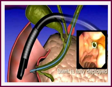
If the bile duct or pancreatic duct is blocked by stones that cannot be removed, or by tumours, the ducts can be made to be passable again by the insertion of flexible metal mesh tubes or stents. These stents are tightly wrapped round catheters that are passed through the Duodenoscope and deployed to bypass the stones or across narrowed segments. Once fully opened up, the tubes are then unblocked and drainage of bile or pancreatic juice is restored.
Conversion of external-internal biliary stent to endoscopic stent ('rendezvous procedure')
Sometimes, due to previous surgery or anatomical variations, ERCP (discussed elsewhere) does not succeed in passing catheters into the bile ducts from the duodenum through the Duodenoscope. In such cases, one option that avoids surgery is a “rendezvous procedure” in which an expert radiologist inserts a tube through the wall of the lower chest into the liver, into a bile duct and delivers a guide wire that is navigated across the obstructed parts of the bile ducts and into the duodenum. A tube is inserted to run from the duodenum and out through the lower chest wall to drain bile. ERCP is subsequently performed and a guide wire is inserted into the duodenum from the lower chest wall, which is then retrieved by a snare loop or wire basket and brought out through the Duodenoscope. In this way, access to the bile ducts is achieved via endoscopy and the problem resolved as per normal ERCP techniques.
Make an Appointment